What's Current in
Life Sciences
Image

Photo Credit
iStock
Six UCSB junior faculty members selected for 2026 NSF CAREER awards
Image

Photo Credit
Matt Perko
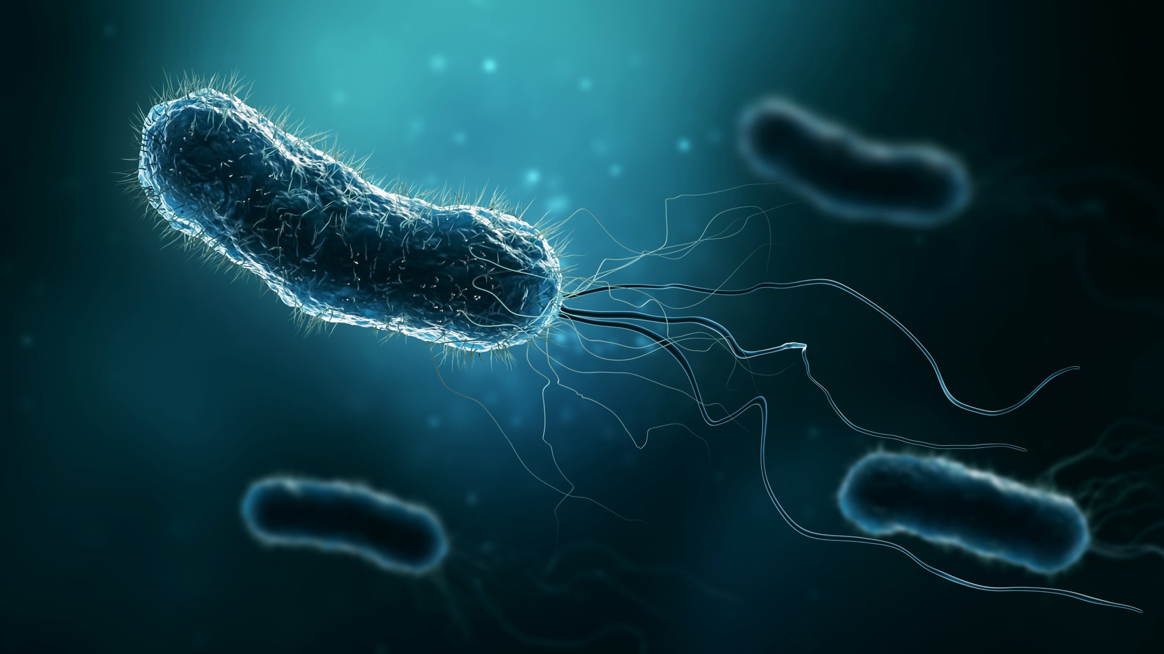
Researchers Mia Raimondi, left, Christopher Hayes and lead author Michael Costello have uncovered how pathogenic Bordetella bacteria adhere to mammalian airways despite their hosts' natural defenses
Image
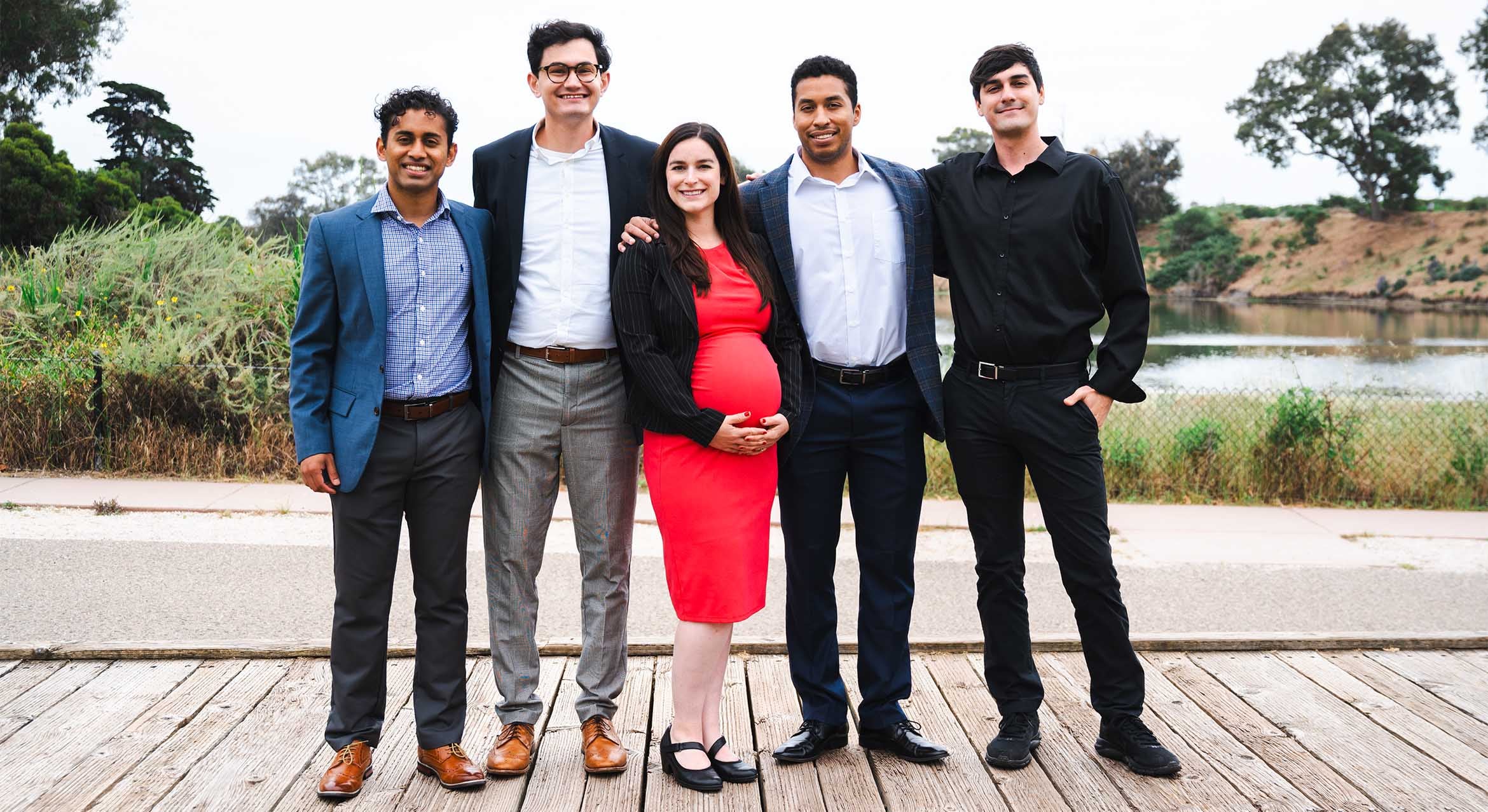
Photo Credit
Courtesy UCSB Engineering
Left to right: Samuel Lobo, Devon Callan, Erica Keane Rivera, Ventura Rivera, Austin Dubose
Image
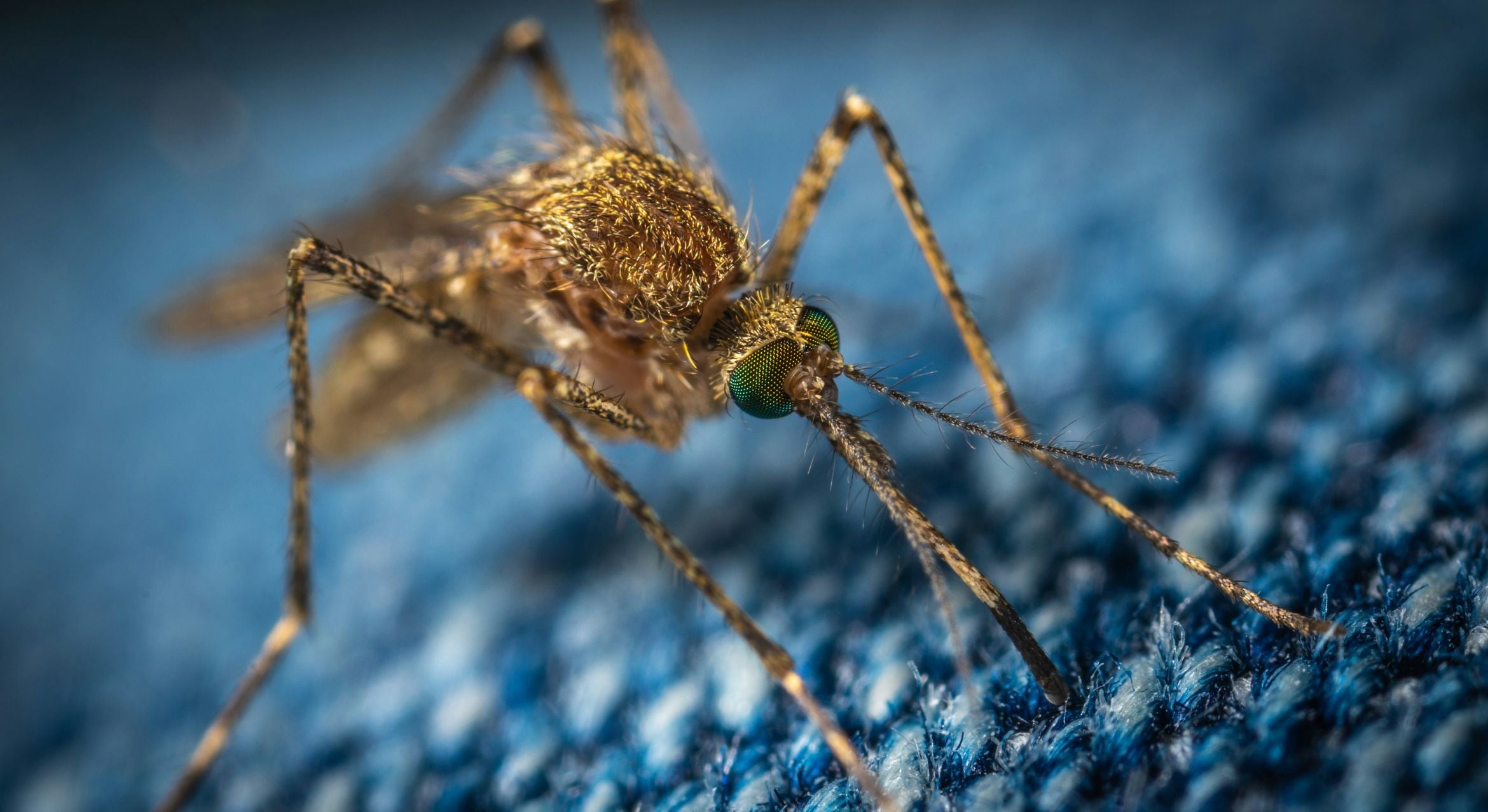
Photo Credit
Photo by Егор Камелев on Unsplash
Finding the right taste to send mosquitoes packing could save hundreds of thousands of lives.
Image
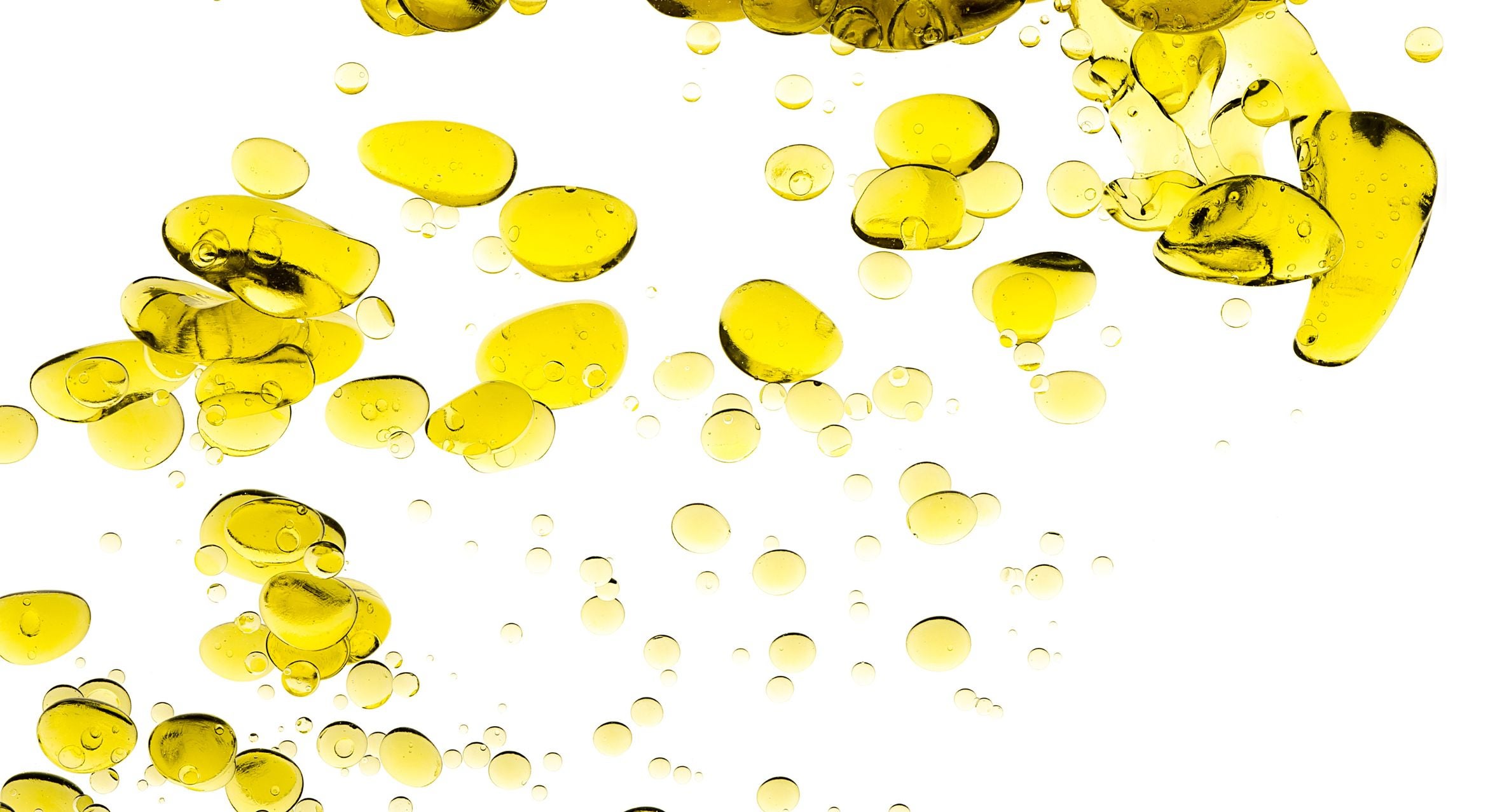
Photo Credit
Carther via iStock
Small, naturally occurring droplets could have accelerated the development of early life.
Image
Microbes are responsible for much of carbon sequestration in the ocean
Image

Photo Credit
Life Science Databases, Creative Commons Attribution-Share Alike 2.1 Japan
The corpus callosum, in red, connects the two hemispheres of the brain together.
Image
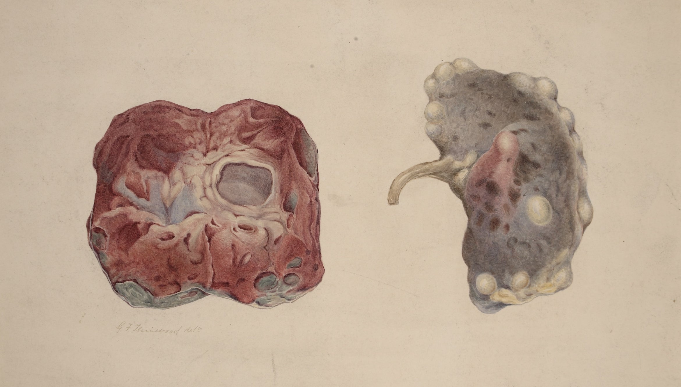
Photo Credit
Teniswood, George Francis, "Polycystic kidney," Barts Health NHS Trust Archives, c1880-1893 CC BY 4.0 <https://creativecommons.org/licenses/by/4.0>, via Wikimedia Commons
Watercolor drawing showing two views of a polycystic kidney. One shows the external surface of the kidney, the other when the organ is bisected. Drawing given to the Museum by Dr. Draper Mackinder, MD, Gainsborough, Lincolnshire
Image
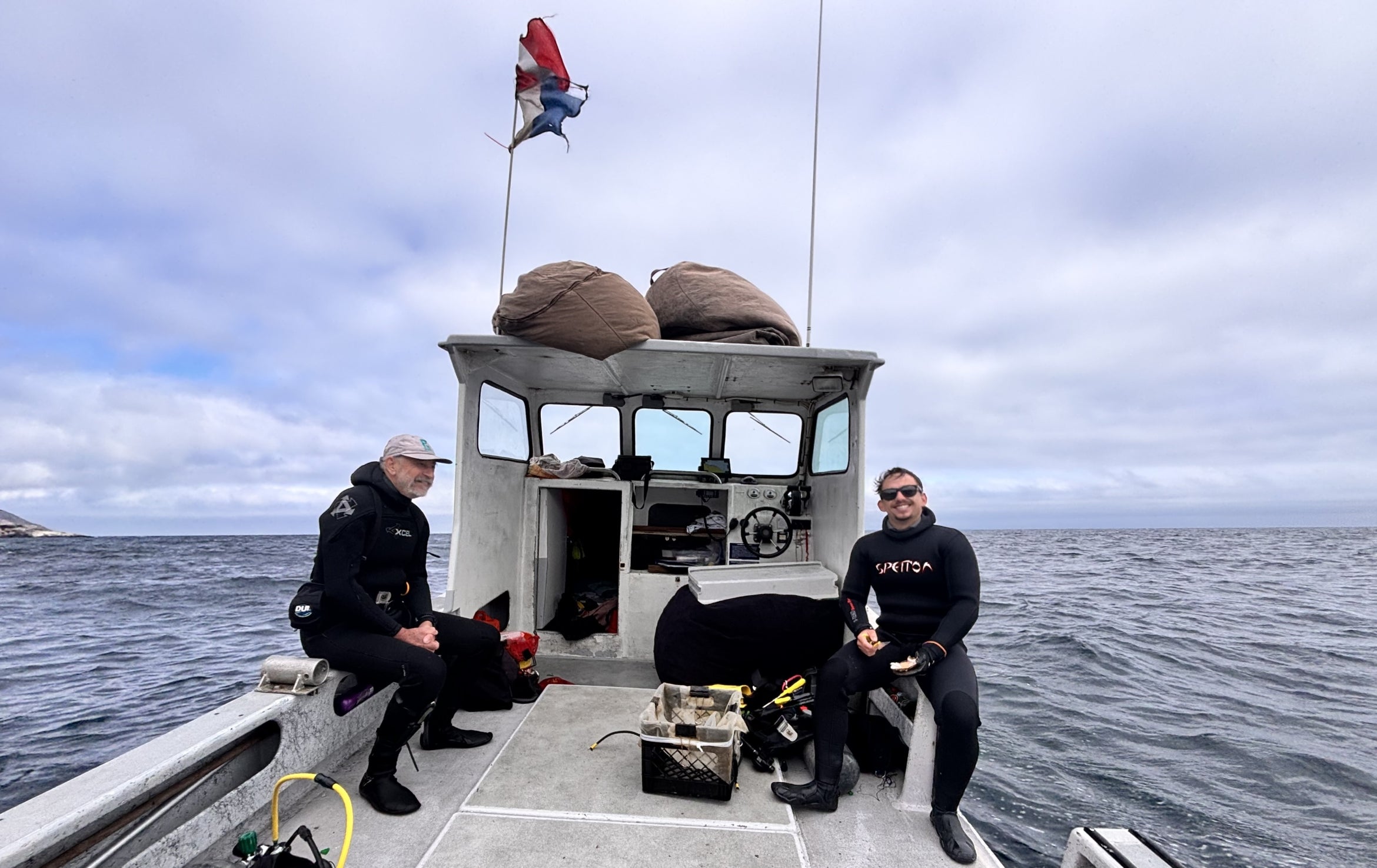
Photo Credit
Courtesy Image
Volunteer taxonomist Gustav Pauly from the Florida Museum of Natural History, left and SBC-LTER lab technician Darrin Ambat on a morning dive to retrieve Autonomous Reef Monitoring Structures from the sea floor
Image

Photo Credit
Jeff Liang
Image
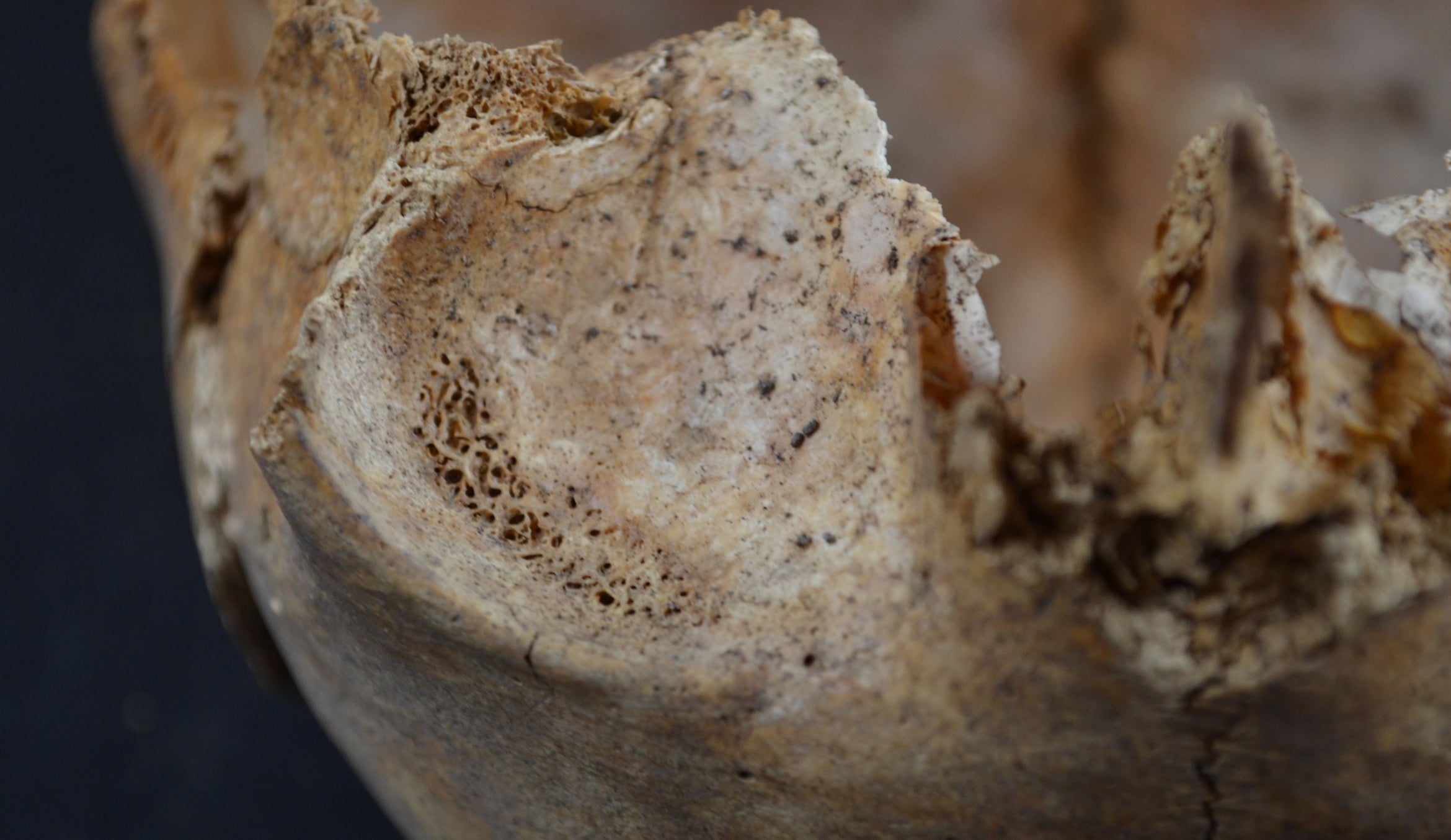
Photo Credit
Dead Men Tell Tales, licensed under Creative Commons Attribution-Share Alike 4.0 International license
Orbital lesions (cribra orbitalia) in a young child from the medieval site of Helgeandsholmen, Stockholm, Sweden