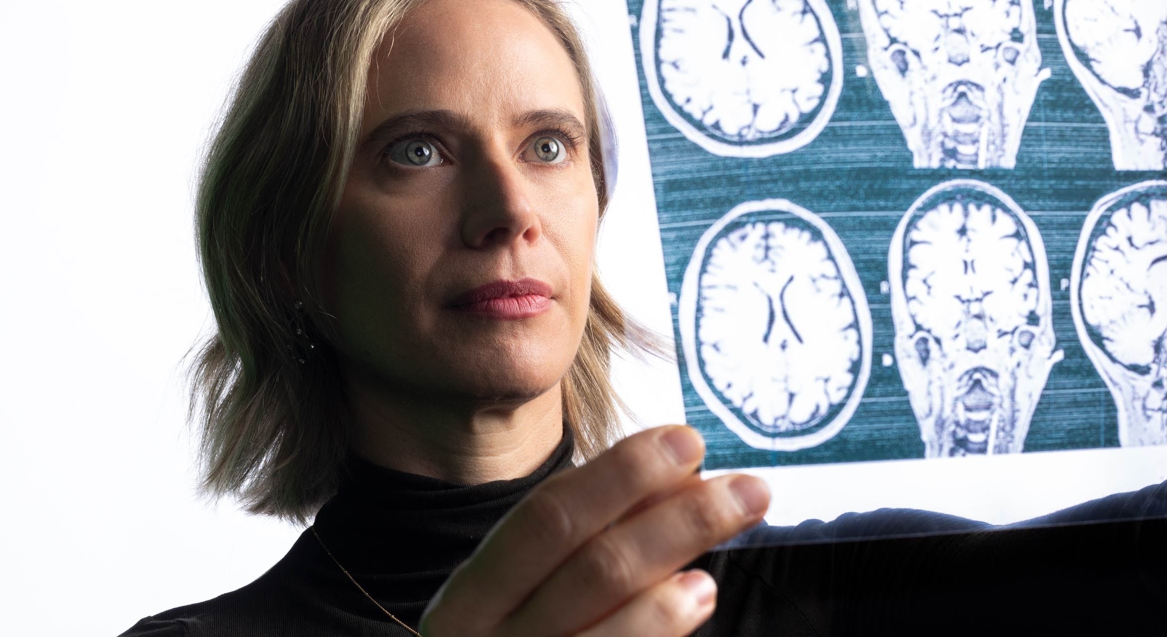
Pregnancy is a transformative time in a person’s life where the body undergoes rapid physiological adaptations to prepare for motherhood — that we all know. What has remained something of a mystery is what the sweeping hormonal shifts brought on by pregnancy are doing to the brain. Researchers in Professor Emily Jacobs’s lab at UC Santa Barbara have shed light on this understudied area with the first-ever map of a human brain over the course of pregnancy.
“We wanted to look at the trajectory of brain changes specifically within the gestational window,” said Laura Pritschet, lead author of a paper just published in Nature Neuroscience. Previous studies had taken snapshots of the brain before and after pregnancy, she said, but never have we witnessed the pregnant brain in the midst of this metamorphosis.
Following one first-time mother, the researchers scanned her brain every few weeks, starting before pregnancy and continuing through two years postpartum. The data, collected in collaboration with Elizabeth Chrastil’s team at UC Irvine, reveal changes in the brain’s gray and white matter across gestation, suggesting that the brain is capable of astonishing neuroplasticity well into adulthood.
Their precision imaging approach allowed them to capture dynamic brain reorganization in the participant in exquisite detail. This approach complements early studies that compared women’s brains pre- and post-pregnancy. The authors noted, “our goal was to fill the gap and understand the neurobiological changes that happen during pregnancy itself.”
Decrease in gray matter, increase in white matter
The most pronounced changes the scientists found as they imaged the subject’s brain over time was a decrease in cortical gray matter volume, the wrinkly outer part of the brain. Gray matter volume decreased as hormone production ramped up during pregnancy. However, a decrease in gray matter volume is not necessarily a bad thing, the scientists emphasized. This change could indicate a “fine-tuning” of brain circuits, not unlike what happens to all young adults as they transition through puberty and their brains become more specialized. Pregnancy likely reflects another period of cortical refinement.
“Laura Pritschet and the study team were a tour de force, conducting a rigorous suite of analyses that generated new insights into the human brain and its incredible capacity for plasticity in adulthood,” Jacobs said.
Less obvious but just as significant, the researchers found prominent increases in white matter, located deeper in the brain and generally responsible for facilitating communication between brain regions. While the decrease in gray matter persisted long after giving birth, the increase in white matter was transient, peaking in the second trimester and returning to pre-pregnancy levels around the time of birth. This type of effect had never been captured previously with before-and-after scans, according to the researchers, allowing for better estimation of just how dynamic the brain can be in a relatively short period of time.

“The maternal brain undergoes a choreographed change across gestation, and we are finally able to see it unfold,” Jacobs said. These changes suggest that the adult brain is capable of undergoing an extended period of neuroplasticity, brain changes that may support behavioral adaptations tied to parenting.
“Eighty-five percent of women experience pregnancy one or more times over their lifetime, and around 140 million women are pregnant every year,” said Pritschet, who hopes to “dispel the dogma” around the fragility of women during pregnancy. She argued that the neuroscience of pregnancy should not be viewed as a niche research topic, as the findings generated through this line of work will “deepen our overall understanding of the human brain, including its aging process.”
The open-access dataset, available online, serves as a jumping-off point for future studies to understand whether the magnitude or pace of these brain changes hold clues about a woman’s risk for postpartum depression, a neurological condition that affects roughly one in five women. “There are now FDA-approved treatments for postpartum depression,” Pritschet said, “but early detection remains elusive. The more we learn about the maternal brain, the better chance we’ll have to provide relief.”
And that is just what the authors have set out to do. With support from the Ann S. Bowers Women’s Brain Health Initiative, directed by Jacobs, their team is building on these early discoveries through the Maternal Brain Project. More women and their partners are being enrolled at UC Santa Barbara, UC Irvine, and through an international collaboration with researchers in Spain.
“Experts in neuroscience, reproductive immunology, proteomics, and AI are joining forces to learn more than ever about the maternal brain,” Jacobs said. “Together, we have an opportunity to tackle some of the most pressing and least understood problems in women’s health.”


