What's Current in
Medicine + Health
Image

Photo Credit
iStock
Glioblastoma tumors are aggressive and difficult to access for treatment
Light-triggered stress response suppresses glioblastoma spread in 3D models
Image

Photo Credit
Matt Perko
Meghan Morrissey
Image

Photo Credit
Courtesy
Film still from the documentary “Walk With Me” about Los Angeles designer Charlie Hess and his journey into early-onset Alzheimer’s.

Image
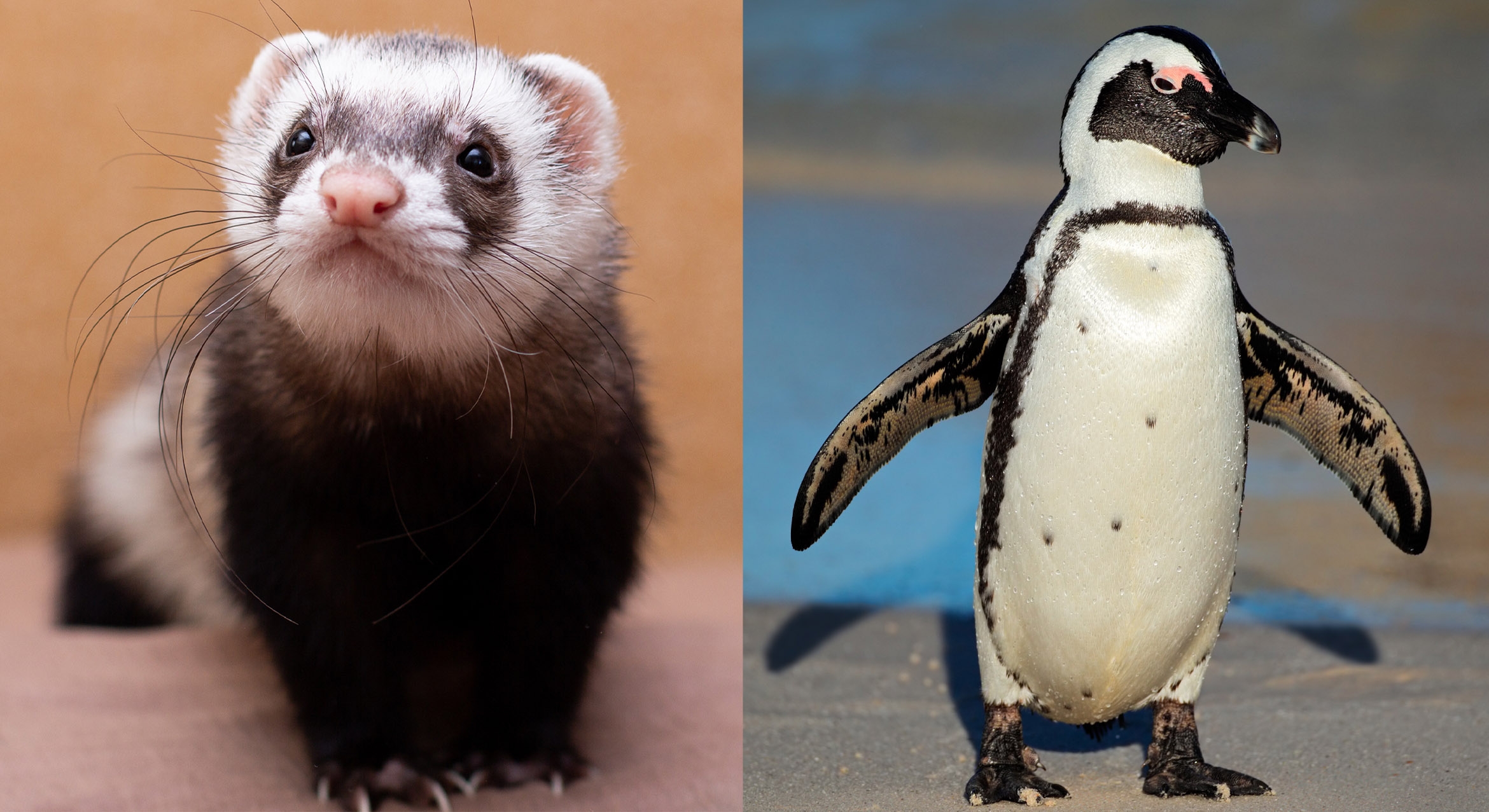
Ferrets are among the vertebrates that get the most tumors, while black footed penguins get the fewest
Image

Photo Credit
Elena Zhukova
Exercising with battle ropes is a popular high-intensity interval training (HIIT) workout
Image

Photo Credit
Matt Perko
Denise Montell
Image

Photo Credit
Matt Perko
Emily Jacobs
Image

Photo Credit
Udra via iStock
Image

Photo Credit
Matt Perko
The small, wearable device is part of a system that monitors drug concentrations and delivers accurate dosages
Image
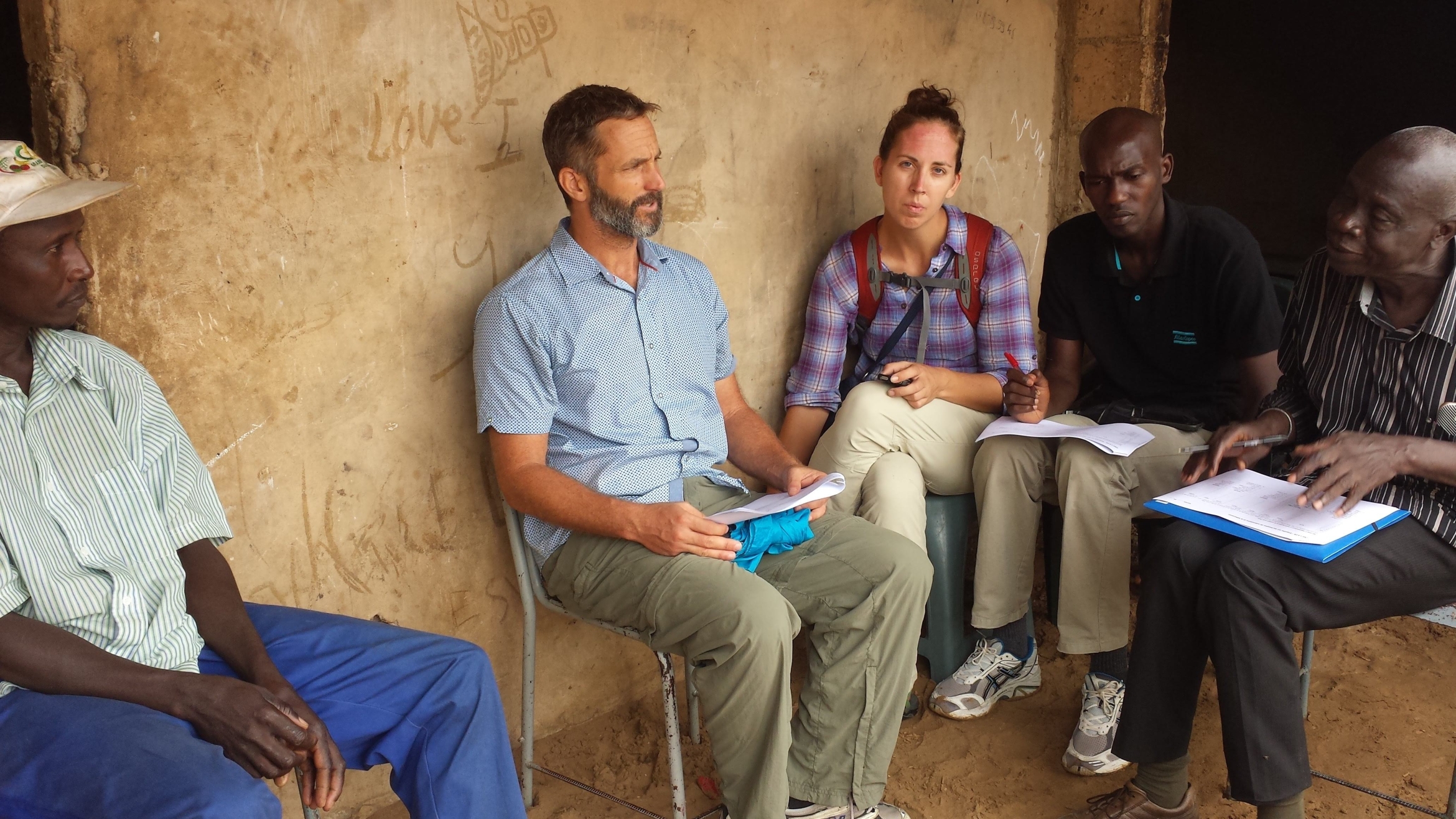
Photo Credit
Courtesy Image
UCSB geographer David López-Carr, center left, and Stanford University health and environmental scientist Andrea Lund, center right, working with Senegalese partners

Image

Photo Credit
Matt Perko
This is a story about proteins, how they malfunction, and what cells do to prevent that.