New microCT machine unites biologists and materials scientists
Share this article
A versatile new device will soon come to campus thanks to an interdepartmental collaboration between biologists and engineers.
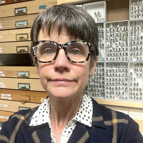
The Nikon microCT scanner is able to image objects from the size of a water bottle to a virus, revealing the internal structure at incredible resolution. Its acquisition was spearheaded by entomologists Katja Seltmann and Madeleine Ostwald at the Cheadle Center for Biodiversity & Ecological Restoration.

“A microCT is important because it allows us to look at internal anatomy and the inside of bee nests as bees are growing,” said Seltmann, the Katherine Esau Director of the Cheadle Center. Most of her group’s previous work has been on external morphology.
Seltmann and Ostwald applied for an equipment grant from the U.S. Department of Agriculture (USDA). The highly selective program fields only one submission per institution each round, so they had to win an internal competition to secure UCSB’s spot. The funding meant they could afford a microCT, but not the most multifunctional model.
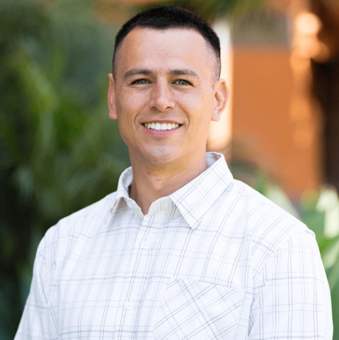
“However, the microCT is useful not just for biology and bee health, but also for anything dealing with morphology, which also includes materials,” she added. The prospect of the new device connected more than 30 faculty across campus, including materials scientist Daniel Oropeza.
The ability to analyze a sample's structure without taking it apart is hugely helpful when studying and designing new structures, Oropeza explained. The microCT will help inform work on additive manufacturing, ceramics, energy storage, bioengineering and more.
The USDA grant will cover about 70% of the cost of the device, with the remaining 30% split between a host of other faculty members and divisions. The College of Engineering; the Division of Mathematical, Life, and Physical Sciences; and the Office of Research each made substantial contributions.
Professor Oropeza pledged money directly from his lab startup funds. His contributions were joined by Marley Dewey (Bioengineering), Paolo Luzzatto-Fegiz (Mechanical Engineering), Jeff Sakamoto (Mechanical Engineering and Materials), and Yangying Zhu (Mechanical Engineering).
The equipment will eventually be available to researchers throughout campus via the Microscopy & Microanalysis Facility once it’s fully operational. “I think the acquisition of the microCT is an amazing example of UCSB's commitment and capacity for cross-campus collaboration,” Oropeza said.
Share this article
What's Current


