Guiding Light

Anyone who has experienced jet lag knows that changing time zones can wreak havoc on our circadian rhythms. Modulated by external cues such as sunlight and temperature, the roughly 24-hour cycles in our physiological processes are extremely sensitive.
Humans aren’t the only creatures whose circadian rhythms are dictated by light. The tiny Drosophila melanogaster — known more commonly as the fruit fly — sets its regular day-and-night-activity cycles in response to light. What’s more, fruit flies also experience jetlag if they experience a sudden shift in the length of one of the day or night cycles.
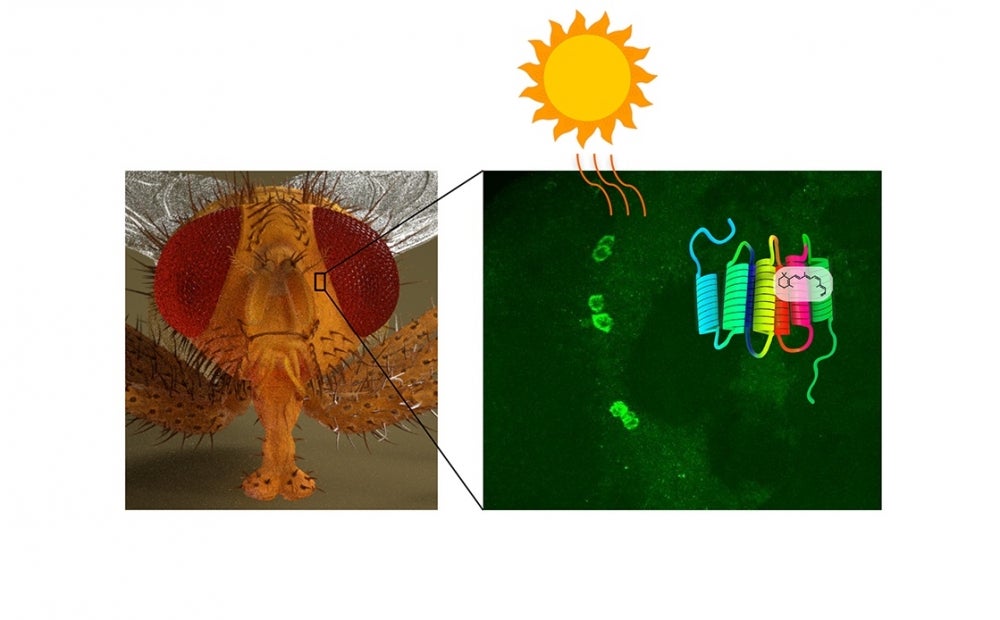
That makes Drosophila so instructive for studying the mechanisms underlying those circadian patterns. Using fruit flies as a model organism, the Craig Montell Lab at UC Santa Barbara has made an unexpected discovery about rhodopsin — a light-sensitive receptor protein common to humans and flies that regulates circadian rhythms through expression in the central brain. The findings are published in the journal Nature.
“Rh7 is the first example of a rhodopsin expressed in the central brain that is important in setting circadian rhythms,” said senior author Craig Montell, the Duggan Professor in UCSB’s Department of Molecular, Cellular and Developmental Biology. “Mammalian opsins are also expressed in many parts of the brain, but their roles are unknown.”
Rhodopsins play well-established roles in image formation in both human and fly eyes. In the fruit fly, six rhodopsins are responsible for the full function of photoreceptor cells in the insects’ eyes. But the role of the seventh, Rh7, was uncharacterized — until now. The Montell group has demonstrated that Rh7 functions as a light sensor that governs daily day-and-night activity cycles.
“The discovery that Rh7 functions in circadian rhythms through expression in the central brain was unexpected,” explained lead author Jinfei Ni, graduate student. “It’s also exciting because this finding expands the roles of these lights sensors, which were originally discovered more than 100 years ago.”
To determine the role of Rh7, Montell, Ni and two collaborators at UC Irvine first confirmed that Rh7 was a real light sensor. They replaced Rh1 — the primary light sensor in the photoreceptor cells of the fly’s compound eye — with Rh7 and found it was a suitable substitute. Next, the researchers established the expression pattern of Rh7 by demonstrating that it was present in the brain’s central pacemaker neurons.
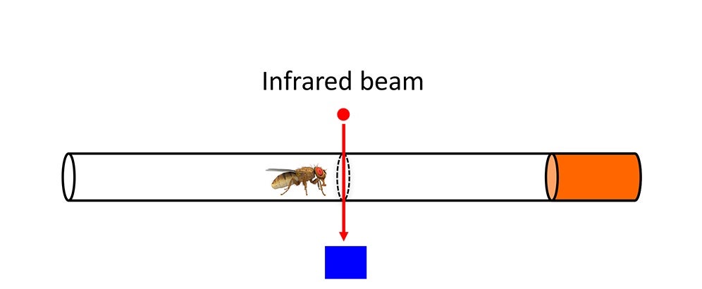
They then performed a series of behavioral studies demonstrating a role for Rh7 in regulating circadian rhythms. In one set of experiments, the scientists maintained the flies on 12-hour day and 12-hour night cycles and then extended one of the day cycles to 20 hours. Normal flies exhibited jet lag but adjusted within a day or two. However, mutant flies missing Rh7 experienced much more severe jetlag that persisted for many days.
Montell posited that the fly’s central pacemaker neurons may correspond to a special type of cell in the mammalian eye. In mammals, retinal ganglion cells (RGCs) receive signals from rods and cones — the light-sensing cells that let us see images — and transmit those signals to the brain via the optic nerve. Only about 1 percent of RGCs are intrinsically photosensitive.
These ipRGCs, which contain melanopsin — a visual pigment more akin to fly visual pigments than those in rods and cones — don’t have roles in image formation. But they are important for the photoentrainment of circadian rhythms. Due to their similar function and molecular features, Montell suggests that the fly’s central pacemaker neurons expressing Rh7 are the equivalent of mammalian ipRGCs.
A light sensor in the central brain works in the fruit fly, because light can pass through the thin cuticle covering the insect’s head. But what could opsins be doing in the mammalian brain? Is it possible that sufficient light penetrates the skull to activate rhodopsins? Montell and colleagues hope additional research will provide an answer.
This work was supported by grants from the National Eye Institute and the National Institute on Deafness and other Communication Disorders.


